Anatomy Of Chest - Boy's chest anatomy, illustration - Stock Image - F018 ... : Anatomy of right side chest pain.. The diaphragm forms the upper surface of the abdomen. This atlas is a comprehensive and affordable learning tool for medical students and residents and especially for radiologists and pneumologists. It provides access to ct images in the axial plane, allowing the user to learn and review the lung anatomy interactively. The chest anatomy includes the pectoralis major, pectoralis minor and the. It is enclosed by the ribs, the vertebral column, and the sternum, or breastbone, and is separated from the abdominal cavity (the body's largest hollow space) by a muscular and membranous partition, the diaphragm.
Female reproductive system cycle fe. The abdomen commonly called the belly is the body space between the thorax. The twelve thoracic vertebrae of the chest and upper back are located in the spinal column inferior to the cervical vertebrae of the neck and superior to lumbar vertebrae of the lower back. Thoracic cavity, also called chest cavity, the second largest hollow space of the body. Muscles of the chest and their functions you have two mighty muscles on both sides of your chest:
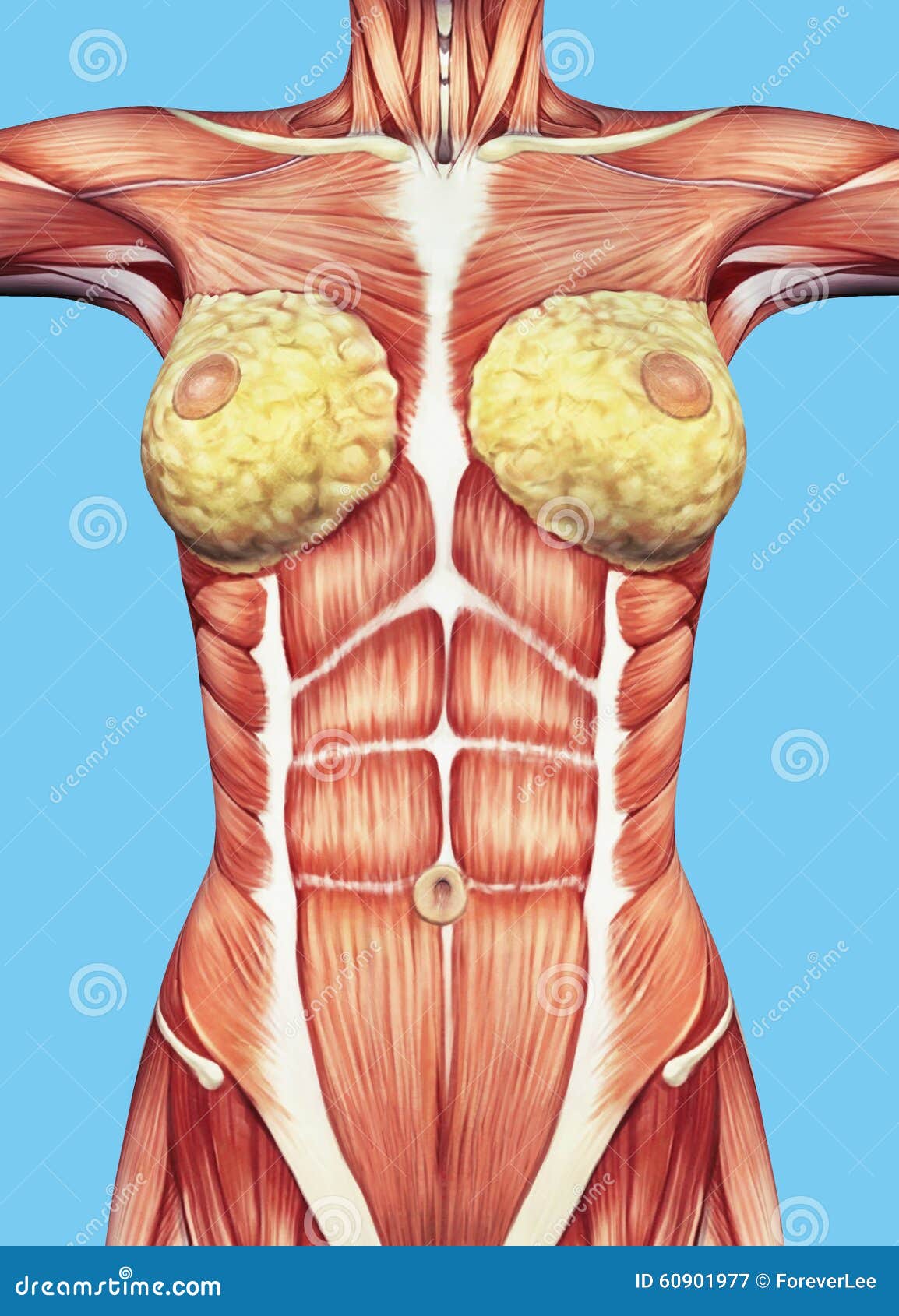
Anatomy Of Female Chest And Torso. Stock Illustration ... from thumbs.dreamstime.com It is enclosed by the ribs, the vertebral column, and the sternum, or breastbone, and is separated from the abdominal cavity (the body's largest hollow space) by a muscular and membranous partition, the diaphragm. The chest is the area of origin for many of the body's systems as it houses organs such as the heart, esophagus, trachea, lungs, and thoracic diaphragm. We think this is the most useful anatomy picture that. This atlas is a comprehensive and affordable learning tool for medical students and residents and especially for radiologists and pneumologists. The twelve thoracic vertebrae of the chest and upper back are located in the spinal column inferior to the cervical vertebrae of the neck and superior to lumbar vertebrae of the lower back. Anatomy of right side chest pain. System respiratory respiratory organs of human body digestive and respiratory system medical chest internal structure of human body medicine body lungs biology intestines stomach anatomy torso human internal. Here, we break down the anatomy of your chest muscles.
This mri chest (thorax) axial cross sectional anatomy tool is absolutely free to use. Several muscles that move the arms, head, and neck have their origins on the sternum. This page provides an overview of the chest muscle group. Principal functions are the protection of internal viscera and an expandable cylinder facilitating variable gas flow into the lungs. Sternocleidomastoid muscle clavicle and ribs anatomy muscle anatomy chest sternocleidomastoid ribs anatomy chest muscles anatomy thorax rib muscles chest muscles chest anatomy illustration. The clavicular head, the sternal head, and the abdominal head. In insects, crustaceans, and the extinct trilobites, the thorax is one of the three main divisions of the creature's body, each of which is in turn composed of multiple segments. Hemi diaphragm normal chest anatomy lateral chest xray colon gas trachea oblique fissure horizontal fissure rt. These heads are important to know because they can be specifically trained through particular movements. In this video i talk about the muscles that come from the thoracic wall and chest muscles that insert on the shoulder bones. The chest or thorax region of the upper body has a number of important organs that reside within it that may present with chest pain if they become compromised in. The chest wall, like other regional anatomy, is a remarkable fusion of form and function. Knowledge of the anatomy of the whole cylinder (ribs, sternum, vertebra, diap …
The chest or thorax region of the upper body has a number of important organs that reside within it that may present with chest pain if they become compromised in. Knowledge of the anatomy of the whole cylinder (ribs, sternum, vertebra, diap … It provides access to ct images in the axial plane, allowing the user to learn and review the lung anatomy interactively. The chest or thorax is the region. See human chest anatomy stock video clips.
Shoulder muscles and chest - human anatomy diagram PDF from i0.wp.com The chest wall is comprised of skin, fat, muscles, and the thoracic skeleton. Browse 6,407 chest anatomy stock photos and images available, or search for human anatomy to find more great stock photos and pictures. It is enclosed by the ribs, the vertebral column, and the sternum, or breastbone, and is separated from the abdominal cavity (the body's largest hollow space) by a muscular and membranous partition, the diaphragm. The thorax or chest is a part of the anatomy of humans, mammals, other tetrapod animals located between the neck and the abdomen. How to view the anatomical labels. This page provides an overview of the chest muscle group. Here, we break down the anatomy of your chest muscles. Learn about each of these muscles, their locations, functional anatomy and exercises for them.
Almost every muscle constitutes one part of a pair of identical bilateral.
Related posts of anatomy of the chest abdominal blood supply. Find subtle abnormalities by using the sihouette sign. This page provides an overview of the chest muscle group. The first step in understanding thorax anatomy is to find out its boundaries. We think this is the most useful anatomy picture that. This atlas is a comprehensive and affordable learning tool for medical students and residents and especially for radiologists and pneumologists. Applied anatomy of the chest wall and mediastinum petros mirilas michael e. The chest anatomy includes the pectoralis major, pectoralis minor and the serratus anterior. Anatomy of the chest and the lungs: How to view the anatomical labels. In a woman these protect the mammary glands. Related images for anatomy of the chest and abdomen male chest anatomy diagram male chest anatomy thorax anatomy. Muscles of the chest and their functions you have two mighty muscles on both sides of your chest:
The first step in understanding thorax anatomy is to find out its boundaries. The pectoralis major and the pectoralis minor, known collectively as your pecs. This page provides an overview of the chest muscle group. These heads are important to know because they can be specifically trained through particular movements. Principal functions are the protection of internal viscera and an expandable cylinder facilitating variable gas flow into the lungs.
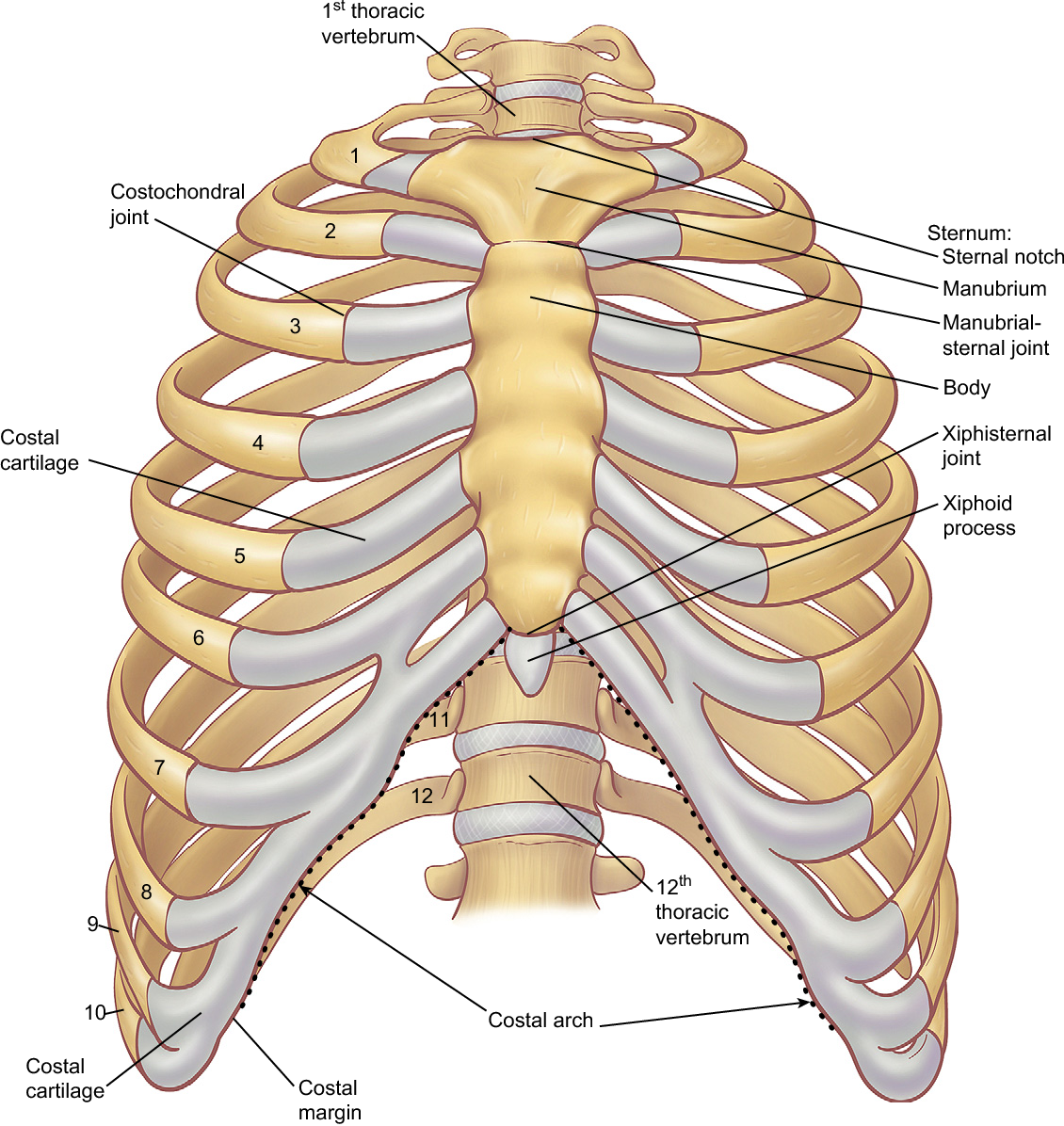
Figure 6 from The anatomy of the ribs and the sternum and ... from ai2-s2-public.s3.amazonaws.com The chest or thorax is the region. Several muscles that move the arms, head, and neck have their origins on the sternum. It is enclosed by the ribs, the vertebral column, and the sternum, or breastbone, and is separated from the abdominal cavity (the body's largest hollow space) by a muscular and membranous partition, the diaphragm. Sternocleidomastoid muscle clavicle and ribs anatomy muscle anatomy chest sternocleidomastoid ribs anatomy chest muscles anatomy thorax rib muscles chest muscles chest anatomy illustration. The chest anatomy includes the pectoralis major, pectoralis minor and the. In insects, crustaceans, and the extinct trilobites, the thorax is one of the three main divisions of the creature's body, each of which is in turn composed of multiple segments. The abdomen commonly called the belly is the body space between the thorax. The epidermis is the outermost layer that provides a protective, waterproof seal over the body.
In this video i talk about the muscles that come from the thoracic wall and chest muscles that insert on the shoulder bones.
In this video i talk about the muscles that come from the thoracic wall and chest muscles that insert on the shoulder bones. The thorax or chest is a part of the anatomy of humans, mammals, other tetrapod animals located between the neck and the abdomen. The diaphragm forms the upper surface of the abdomen. See chest anatomy stock video clips. Computed tomography (ct) of the chest can detect pathology that may not show up on a conventional chest radiograph(1). This thoracic and pulmonary anatomy tool is especially designed for students of anatomy (medical and paramedical studies). See human chest anatomy stock video clips. Skandalakis chest wall embryogenesis the muscles of the chest develop from the somites found in the mesoderm. The chest is the area of origin for many of the body's systems as it houses organs such as the heart, esophagus, trachea, lungs, and thoracic diaphragm. Chest a man's chest — like the rest of his body — is covered with skin that has two layers. About the 6th week, the somites differentiate into the sclerotomes and the dermatomyotomes. Principal functions are the protection of internal viscera and an expandable cylinder facilitating variable gas flow into the lungs. Thoracic cavity, also called chest cavity, the second largest hollow space of the body.